Mauritanian Anatomy Laboratory Thoracic Atlas
Launch Mauritanian Anatomy Laboratory Thoracic Atlas
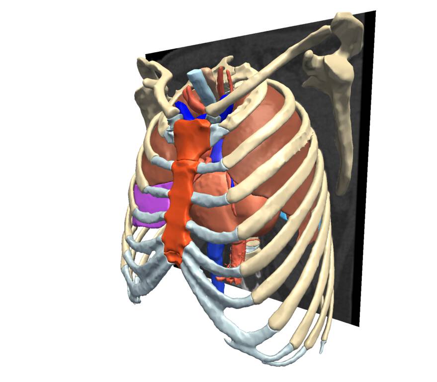
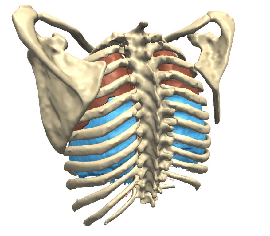
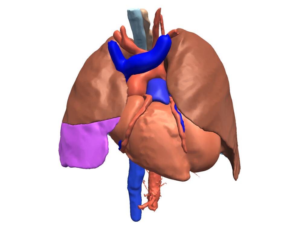
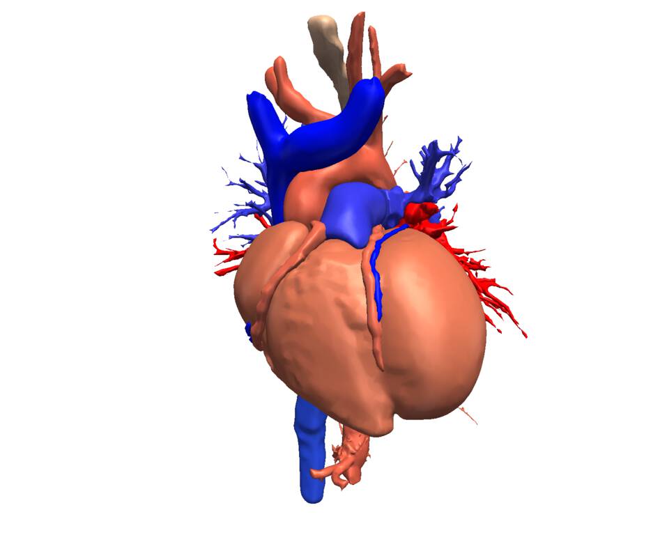
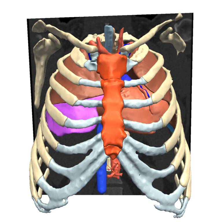
Derived from a high quality CT scan, this atlas of the thorax from the Mauritanian Anatomy Laboratory (MAL) includes thoracic skeletal system, the respiratory system, the cardiovascular system, and the esophagus.
Description
This Thorax Atlas is provided by the team of Mauritanian Anatomy Laboratory (MAL) of the Faculty of Medicine (FM) at University of Nouakchott Al Aasriya in Mauritania. Its reconstruction was created using the 3D Slicer platform. It includes the skeletal system of the thorax, the respiratory system, the cardiovascular system, and the esophagus. The current atlas version was derived from an original DICOM computed tomography (CT) scan, segmented using 3D Slicer’s reonstruction techniques including semi-automated and manual image segmentation and modules such as Models, Volumes, Segmentation, Segment Statistics Segment Editor (+Extra Effects).
Data of a patient 38 year old male was acquired for this atlas by the Radiology Department at the National Cardiovascular Centre (CNC). Imaging was performed on a Toshiba prime 80 CT scanner, using a multi-array head coil. Labels were generated using free surfer automatic parcellation followed by manual segmentation of most structures.
Authors
- Haythem Guermazi (Undergraduate Medical Student)
- Ahmedou Moulaye IDRISS (Head of Anatomy Lab. 1)
- Tfeil Yahya (Head of Anatomy Lab. 2)
Institutions
-
Anatomy Lab, Faculty of Medicine, University of Nouakchott Al Aasriya (UNA), Nouakchot, Mauritania.
-
Cardiovascular National Centre, Nouakchott - Mauritania
Other contributors
- Khaled Boye (Head of Cardiovascular Surgery Department CNC)
- MACBiolDi Project, ULPGC, Canary Islands, Spain.
Release date
January 2021
Keywords
thorax, spine, cardiovascular system, pulmonary system
Sponsors and funding
- P41 RR013218/RR/NCRR NIH HHS/United States
- P41 EB015902/EB/NIBIB NIH HHS/United States
- This work is funded as part of the Neuroimaging Analysis Center, grant number P41 RR013218, by the NIH’s National Center for Research Resources (NCRR) grant number P41 EB015902, by the NIH’s National Institute of Biomedical Imaging and Bioengineering (NIBIB) and the Google Faculty Research Award.
License
Download
https://www.openanatomy.org/atlases/macbioidi/mauritania/thorax-2020-11-15.zip
Other images
